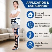
1. Classification of common traction techniques in orthopedics: (1) According to the location of traction, it is divided into spinal traction and limb joint traction. Spinal traction is further divided into cervical traction and lumbar traction. (2) According to the traction power, it is divided into manual traction, mechanical traction, and electric traction
Both fracture reduction and surgical internal fixation should be followed by immobilization and rest. Use triangular scarves or low-temperature board supports to externally fix functional positions for repair purposes. Wrists and fingers can be used for flexion and extension exercises.
3-4 weeks, patients with internal fixation can practice shrugging and pectoralis major and latissimus dorsi muscle groups; Practice the flexion and extension function of the elbow joint 10 times per group, every 20-30 seconds, for 2-3 groups.
The deltoid muscle contracts without resistance, and it is important to avoid excessive abduction angles. Do not engage in shoulder abduction or elbow joint extension and flexion exercises.
From 5-8 weeks, gradually increase the intensity of the above exercises, and appropriately increase resistance training and upper arm internal and external rotation exercises. External fixation can be removed as appropriate. Perform weightless pendulum arc motion training on the shoulder joint.
After 8 weeks, perform shoulder joint abduction exercises using ribbed shoulder ladders, hanging pulleys, wall tensioners, and other equipment. If there is movement disorder in the shoulder joint, rehabilitation treatment is performed through joint pulling surgery, with the same technique as shoulder dislocation.
Fractures that require manual reduction can be treated with shortwave therapy after 1 week, followed by local ultrasound treatment after 2 weeks, using the same method as before.
4. Common treatment methods for hand nerve injury repair: (1) physical factor therapy (2) exercise therapy ① massage and passive movement ② "impulse transmission" training ③ muscle strength training ④ sensory training ⑤ electromyographic biofeedback (EMGBF) (3) occupational therapy (4) brace application (5) coordination ability training (6) psychological therapy (7) ADL training 5. List 5 rehabilitation training methods for lower limb fractures: quadriceps isometric contraction exercise CPM training, standing and walking, squatting straight leg lifting training, proprioceptive and power cycling training 6. Common complications after total hip arthroplasty: ① postoperative dislocation ② deep vein thrombosis ③ pain ④ prosthesis loosening ⑤ ectopic ossification 7. Total knee arthroplasty (TKA) R) The ultimate success depends on five aspects: ① selection of case indications ② prosthesis design ③ prosthesis materials ④ surgical techniques ⑤ implementation of postoperative rehabilitation techniques 8. Clinical staging of shoulder periarthritis: 1. Acute phase: lasting 10-36 weeks, mainly characterized by severe pain and tenderness around the shoulder joint, accompanied by muscle spasms and limited shoulder joint movement. 2. Freezing period: lasting for 4-12 months. Pain symptoms are relieved and tenderness is still widespread. Shoulder joint contractures are severely restricted, presenting a "frozen" state. 3. Recovery period: lasting for 5-26 months. Pain gradually disappears, shoulder joint contractures and adhesions gradually disappear, and shoulder joint activity gradually recovers.
1. Describe the rehabilitation steps for pelvic and acetabular fractures: 1. Within 2 weeks after early finger injury. The purpose of rehabilitation training is to promote blood circulation in the affected limb, in order to facilitate swelling reduction and fixation. 2. Clinical healing from 2 weeks after mid-term finger injury to fracture. It should gradually shift from passive activity to active activity. 3. Late stage refers to when the fracture has reached clinical healing or external fixation has been removed. The main purpose is to restore the range of motion of affected joints, enhance muscle strength, and restore normal limb function. 2. Elaborate on the specific content of rehabilitation in the later stage of spinal cord injury: generally refers to 8-12 weeks after onset. The target evaluation method for this period is basically the same as early training, which focuses on obtaining posture control and balance ability. 1. Main content and sequence of functional exercise for quadriplegic patients: 1. Bed rest training, 2. Sitting training, 2. Main content and sequence of functional exercise for paraplegic patients: 1. Lying training, 2. Turning exercises, 3. From lying to sitting exercises, 4. Sitting balance exercises, 5. Sitting movement exercises, 6. Diagonal bed standing treatment, 7. Balance bar exercises, 8. Walking training (1. Four point step training, 2. Swing to step training, 3. Swing over step training) 9 Standing exercise 10 Crutch exercise 11 Weight reduction training 3 Wheelchair exercise 1 Transfer training 2 Walking in and out of the wheelchair with crutches 3 Balance exercise 4 Walking up and down steps 5 Functional activities on the wheelchair (1 Picking up objects from the floor 2 Reaching the foot pedal with hands 3 Decompression action) 4 Use of orthosis 5 Physical factor treatment 1 Functional electrical stimulation 2 Other physical factor treatment: Ultrasonic low, medium and high frequency electrotherapy Magneto optic wax therapy Transcranial magnetic stimulation treatment 6 Hyperbaric oxygen treatment 7 Occupational treatment 8 Traditional Chinese medicine therapy 1 Massage 2 acupuncture and moxibustion 9 Complications and prevention and treatment: Complications may occur at all stages, which are important factors leading to patient death, so comprehensive prevention and treatment measures should be taken, especially strengthening rehabilitation function training Common complications: compression Sore respiratory system, urinary system, cardiovascular system and metabolic disorder.
1. The concept and application of prostheses: prostheses are artificial limbs that compensate for human limb defects and compensate for limb functions. They can be divided into temporary prostheses and formal prostheses. Clinical prostheses: prostheses installed after the healing of the residual incision generally have good wound healing 2-3 weeks after amputation, and can be installed after removing the thread. They are usually worn for 2-3 months to reduce swelling, prevent contractures, accelerate shaping, and provide reference for the installation of formal prostheses. 2. Postoperative immediate installation of prostheses: Temporary prostheses installed on the operating table immediately after amputation surgery are not commonly used to reduce phantom limb pain and complications, promote wound healing, and shorten bed rest time. 3. Formal prosthetics: Can be used for long-term use by patients to improve their independent living and activity abilities through training such as balance training, walking training, etc. 2. Orthopedic rehabilitation nursing content:
Prevention and treatment of adverse reactions caused by long-term bed rest (such as early activity to prevent disuse syndrome, regular turning over to prevent pressure ulcers, encouraging patients to actively engage in various activities, prevention and treatment of urinary and fecal dysfunction, etc.)
Guide patients to independently engage in daily activities such as dressing, eating, and washing up
Cooperate with training patients' limb movement functions (such as sitting, standing, walking, etc.)
3. Principles for Rehabilitation Treatment of Shoulder and Acromioclavicular Joint Dislocation:
After reduction, the shoulder joint should be braked. In a fixed posture of the shoulder joint, instruct the finger, wrist, and elbow extension and flexion function training. Active resistance training can be carried out to prevent muscle atrophy and joint contraction. Local cold therapy can prevent swelling, bleeding, and reduce pain.
After 3 days, under the above training, remove the cold therapy and perform the following locally:
(1) Ultra short wave therapy involves bipolar opposition at the affected area, with no or minimal heat, for 10-15 minutes each time, once a day, for a course of 10 days.
(2) Ultrasonic therapy, local contact movement method, 15 to 20 minutes each time, once a day, with 10 sessions per course of treatment.
In the third week, one can actively engage in shoulder movements, including front and back movements
The inward and outward movements should be gentle, slow, and not too forceful.
From the 4th to the 6th week, after removing the fixed object: (1) anterior posterior and internal external swing of the shoulder joint, active shoulder abduction, extension, and internal external rotation movements, assisted resistance and passive joint function training. (2) The application of gymnastics sticks, high hanging pulleys, dumbbells and other equipment can improve joint mobility and muscle strength. (3) Practice the functions of shoulder stairs and ribs. (4) Wall brace or rubber band training to enhance shoulder joint mobility and shoulder strap muscle strength. (5) Shoulder joints with limited range of motion
Use joint mobilization surgery. The patient is in a supine position, assisted by auxiliary personnel to fix the scapula. The therapist applies tension (separation) to the joint at any position on the glenoid plane, with a strength of grade III, limited to the patient's tolerance of painless or mild pain. The next day, the patient is asked if there are any adverse reactions such as local pain or swelling, and the technique and intensity are adjusted in a timely manner until the joint function returns to the optimal range of motion. 4. Peripheral nerve injury can be classified according to Seddon's method as follows: nerve loss (Grade I injury), axonal rupture (Grade II injury), nerve rupture (Grade III, IV, V injury). Rehabilitation assessment for multiple hand injuries includes:
(1) Joint range of motion assessment (II) Muscle strength assessment (III) Sensory assessment (IV) Hand function assessment (V) Activities of daily living assessment 6. Rehabilitation assessment for lower limb fractures:
1. Length and circumference: 2. Muscle strength. 3. Joint mobility. 4. Gait. 5. Lower limb function assessment 6 Neurological function 7 Pain sensation function. 8. Balance ability. 9. Daily life ability 10 Fracture healing status
7. Principles of spinal cord injury functional training: 1. Early stage 2. Gradual progression 3. Starting from functional needs 4. Strength and endurance training 8. The most common complications after total shoulder arthroplasty: 1. Loose prosthesis 2. shoulder joint instability 3. infection 4. rotator cuff injury 5. ectopic ossification 1. Definition and content of early rehabilitation for spinal cord injury: 1 Early stage: acute bedridden period
Prevention of pressure ulcers, spasms, and respiratory problems is usually most important within 6-8 weeks after onset of the disease
1. Respiratory care: Deep breathing techniques, vibration, intermittent positive pressure breathing, and cough assistance techniques can all be applied in a timely manner. 2. Actively or passively move joints,. 3. Maintain joint function position. 4. Regularly switch positions to prevent pressure ulcers. 5. Selective muscle strength training. 6. Perform upright movement after X-ray examination shows that the fracture has stabilized or has sufficient internal fixation. (To prevent orthostatic hypotension, gradually adapt using a shaker or inclined bed. Use elastic bandages or elastic socks to accelerate lower limb venous lymphatic return.) 7. Daily activity training. 2. Describe the rehabilitation assessment content of OA: 1. Pain assessment: Use visual simulation scoring index results to judge: 0-3 mild pain 4-7 moderate pain 8-10 severe pain 2. Measurement of limb circumference and joint circumference 3. Muscle strength assessment (1) Knee OA quadriceps and hamstring muscle strength (2) Hip OA hip flexion and extensor muscle strength, hip adduction and abduction muscle strength, hip internal and external rotation muscle strength (3) Hand OA metacarpophalangeal joint, proximal interphalangeal joint, distal interphalangeal joint muscle strength Muscle related to joint flexion and extension
Measurement of grip strength of adductor and abductor muscles (4) Spinal joint OA mainly detects muscle strength related to cervical and lumbar flexion and extension activities. 4. Measurement of joint range of motion: The purpose of evaluation is to understand the degree of joint activity restriction in the affected joints and determine whether it affects daily life activities. 5, Hand function assessment 6, lower limb function assessment Harris hip joint function assessment standard HSS knee joint assessment system Maryland foot function assessment standard 7, daily living activity ability assessment Joint dysfunction impact assessment on ADL Stewart physical activity ability assessment 8, quality of life assessment can be assessed using the Meenan Joint Impact Scale.